 "
"
Team:Harvard/Laser
From 2009.igem.org

Characterization/***********************************************************************************************************************/
Phycocyanobilin. PhyB function requires the covalent attachment of the prosthetic group phycocyanobilin, or PCB. Normally, plants and algae produce this prosthetic group, phycocyanobilin, or PCB, which attaches to the PhyB auto-catalytically. However, since yeast cells don’t normally express Phytochrome B, they also do not have the enzymes required to create PCB. Thus PCB had to be added to cultures for PhyB to function. Because PCB is not commercially available, we had to extract PCB from Spirulina algae, obtained from the health food store The Vitamin Shoppe.
 , ,In this timecourse experiment the x-axis is time in hours, and the y-axis is culture density, as assayed using spectophotometry. The concentrations were measured at 600 nm. Cultures all began with the same initial cell concentration, hence the only variable between the PCB containing culture and the DMSO containing control culture was the presence of PCB. All absorbances were normalized to the absorbance of medium without cells. Each data point is the average of three replicates. We found that the cultures containing PCB actually grew better. From this data we concluded that our crude extract had no toxic effects, and thus decided that we would proceed with the experiments without pursuing further purification of the crude PCB. Inital Screen for Functional Colonies Once we created the necessary plasmids and fusion protein constructs, the first order of business was to find out if the PhyB/PIF3 two-hybrid system worked in our yeast. We began with assays of transformed cells grown on solid medium. The strain of yeast we used were Y190 (MATa, ura3-52, his3-D200, lys2-801, ade2-101, trp1-901, leu2-3, 112, gal4D, gal80D, URA3::GAL1UAS-). Because the cells are Trp- and Leu-, we were able to select for only colonies containing our plasmids (which contain the genes for Trp and Leu synthesis) by growing the cells on Trp-/Leu- plates. Those plates were then exposed to red light from a fluorescent light covered with a red filter (Roscolux) overnight. The filter lift assay was conducted. Nitrocellulose filters were laid on top of the plates allowing cells to adhere to them, and the filters were then dipped in liquid nitrogen to lyse the cells. The nitrocellulose filters were then allowed to sit in buffer containing X-gal, which turns blue when broken down by beta-galactosidase, thus indicating the presence of beta-gal. As is visible below, there were a number of colonies that demonstrated beta-gal production only in the presence of light and PCB. We concluded that these cell lines contained the desired functional PhyB/PIF3 two hybrid system. Screen assay—petri dishes For colony lifts from solid agar, nitrocellulose filters were used. After cutting an appropriately sized filter to fit the surface of a petri plate, nitrocellulose was laid on the agar surface and then removed using tweezers. The filters were then placed onto an aluminum sheet that was floated on liquid nitrogen, for approx. 20 seconds. The filters were then immersed in liquid nitrogen for 2 seconds, thawed at room temperature. Filter lifts were then placed cell side up onto pads of Whatman #1 filter paper, in empty petri-plates, that had been soaked with Z-buffer solution containing 2-mercaptoethanol and X-gal.The filter lifts were then incubated at 30 C for 30min to overnight, until the development of a blue precipitate was clearly visible. For the initial screen, after transformation with plasmids containing the PhyB-DBD and Pif3-AD, plates were pre-incubated overnight , in the dark, at 30 deg. C with 25 uM PCB or DMSO only. After replica plating onto similar media, the plates were then incubated overnight, under red light. Positive colonies were then picked that expressed beta-galactosidase only under red-light induction. These were then re-streaked onto the same leu- trp- selection media and rescreened under overnight growth in red light, in the presence of PCB. 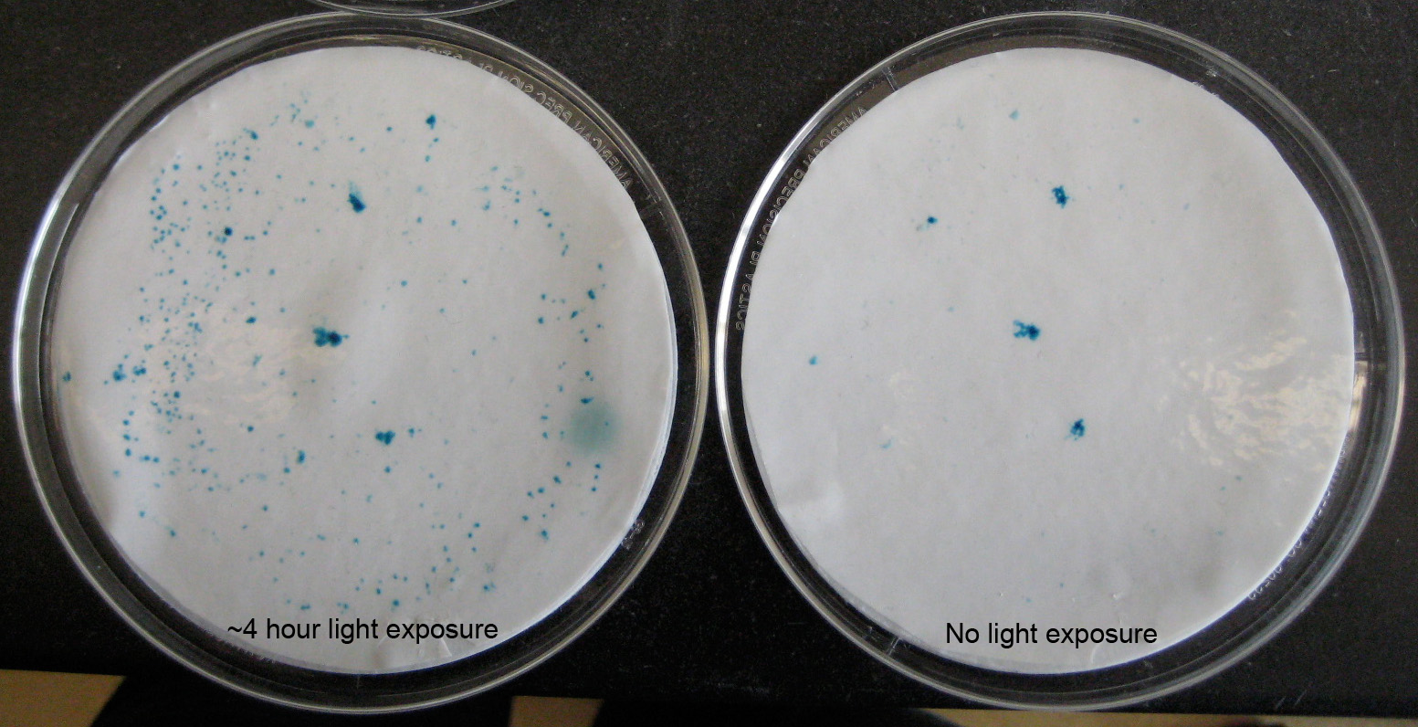 , , , ,Characterization assays—biodot and cotton applicators (“q-tips”) A similar approach was used in the semi-quantitative assays. Alternatively, yeast cells from liquid culture were immobilized onto nitrocellulose filter sheets using a Bio-Rad “Bio-Dot” apparatus connected to a vacuum manifold. The immobilized cells were lysed using liquid nitrogen and incubated with x-gal containing buffer as outlined above. For the analysis of beta-galactosidase expression in cells grown in 96 well micro-titer dishes, cotton applicators were used to remove cells from each well and then immersed directly into liquid nitrogen. After thawing at room temperature, they were then incubated for ~30-overnight in wells containing 150 uL of X-gal buffer.  , ,PCB Concentration Assay For the PCB concentration assay, Y190 yeast cells containing the PhyB-DBD/Pif3-AD system were grown in selective leu- trp- liquid media to a density of 1 X10⁶ cells/ml and 1 X 10⁴ cells were plated in each well of a 96 well microtiter plate, containing 100 uL of solid media containing differing concentrations of PCB: 10 uM, 25 uM and 50 uM or equivalent volumes of DMSO carrier. All tested concentrations of PCB proved effective in inducing lacZ expression.  , ,For the laser pulse duration assay, yeast cells were grown and plated to the same density as mentioned for the PCB concentration assay. Cells were then treated to several pulse durations of light using a ~650nm wavelength, <3mW laser. The pulse durations used were ~0.5 seconds, 10 seconds and 30 seconds. All laser pulse durations were successful in inducing reporter expression, with the highest induction occurring at 10 seconds.  , ,Incubation Time After Light Induction For the reporter expression time course analysis, cells were grown in liquid culture in the same conditions as mentioned for the PCB concentration experiment mentioned above. 150 uL of cells in liquid media were then transferred into individual wells in a 96 well microtiter dish. Cells were then pulsed for 10 seconds using a laser, incubated for either 30 minutes or 1 hour in the dark and then the individual wells of cells were transferred onto nitrocellulose filter paper using a “bio-dot” apparatus connected to a vacuum. Significantly higher expression levels of beta-galactosidase were detected after 1 hour of growth, post laser induction vs. 30 minutes.  , ,Toubleshooting Problems with cell age In our experiments, we found that whether or not the assay worked is highly dependent on cell age. In the following experiment, we grew cells in liquid medium with PCB in the dark (or DMSO for control), then pipetted an aliquot of cells into each well of a 96 well plate. Each well was exposed to red laser light for the indicated amount of time, and then covered and allowed to incubate for 3 hours. After the incubation period the wells were swabbed with q-tips, which were then dipped in liquid nitrogen to lyse the cells, and then submerged in x-gal solution. Q-tips with cells producing beta gal turned blue. In this assay, we found that the older cells had higher background in the negative control than the younger cells. This is probably attributable to the cell line we used, Y190. This cell line contains both beta-galactosidase and histidine reporters under the control of the Gal promoter. Because the cells do not typically produce and mutation would give those mutant cells a metabolic advantage. Any mutations in that would upregulate His production could also lead to an increase in constitutive beta-gal expression. We attempted to reduce this effect by supplementing our media and plates with additional His, but this hypothesis remains untested and further exploration is needed. Problems with concentration We also experienced problems with cell concentration: If cell concentration was too high, we saw significant positive background. This is probably due to low levels of production of beta gal in the absence of induction, which is magnified as cell concentration increases, resulting in high positive background on controls if the cells used are too concentrated. Although it is preferable to not allow the cells to overgrow, we have evidence that dilution of a culture works to help reduce positive background. In this assay, we exposed cells to light or dark according to the following diagram, doing sequential dilutions to examine the effects of cell concentration. Although at the highest concentration there was significant background on the negative control, at a 10 fold dilution, the background is reduced significantly. Further dilutions are less effective because the blue becomes too difficult to see.  , , , , , , |
TeamProjects
Lab Notebook |