| Component
| Description
| Part/Accession #
| Base Pairs
| Plasmid
| Resistance
| Well
|
| RBS
| Ribosomal Binding Site
| [http://partsregistry.org/Part:BBa_B0034 BBa_B0034]
| 12
| pSB1A2
| Ampicillin
| plate 1, 2M
|
| Red Light Sensor
| description
| [http://partsregistry.org/Part:BBa_I15010 BBa_I15010]
| 2,238
| pSB2K3
| Kanamycin
| N/A
|
| OmpR (E. coli)
| description
| [http://www.ncbi.nlm.nih.gov/sites/entrez?Db=gene&Cmd=retrieve&dopt=full_report&list_uids=947913&log$=databasead&logdbfrom=protein NP_417864.1]
| 720
| pSB1T3
| Tetracycline
| N/A
|
| Terminator
| Stops Transcription
| [http://partsregistry.org/Part:BBa_B0015 BBa_B0015]
| 129
| pSB1AK3
| Ampicillin
and Kanamycin
| plate 1, 23L
|
| OmpR + Terminator
| description
| sequence
| 916
| pany-amp
| Ampicillin
| synthesized
|
| OmpC promoter
| description
| [http://partsregistry.org/Part:BBa_R0082 BBa_R0082]
| 108
| pSB1A2
| Ampicillin
| plate 1, 16K
|
| puc B/A
| description
| sequence
| 375
| pSB1A3
| Ampicillin
| N/A
|
| puc B
| description
| [http://www.ncbi.nlm.nih.gov/sites/entrez?Db=gene&Cmd=retrieve&dopt=full_report&list_uids=3719170&log$=databasead&logdbfrom=nuccore YP_353390]
| 156
| ?
| ?
| N/A
|
| puc A
| description
| [http://www.ncbi.nlm.nih.gov/sites/entrez?Db=gene&Cmd=retrieve&dopt=full_report&list_uids=3719171&log$=databasead&logdbfrom=nuccore YP_353391]
| 165
| ?
| ?
| N/A
|
| OmpC promoter+BA
| description
| sequence
| 539
| pany-kana
| Kanamycin
| synthesized
|
| Light Response System
| description
| [http://partsregistry.org/Part:BBa_M30109 BBa_M30109]
| 4,333
| ?
| Ampicillin
| N/A
|
| TetR repressible
| description
| [http://partsregistry.org/Part:BBa_J13002 BBa_J13002]
| 74
| pSB1A2
| Ampicillin
| plate 1, 13B
|
| Green Flourescent Protein
| Marker for successful transformation
| [http://partsregistry.org/Part:BBa_E0240 BBa_E0240]
| 976
| pSB1A2
| Ampicillin
| plate 1, 12M
|
Back To Top
Characterization
 Volver Arriba
Modeling
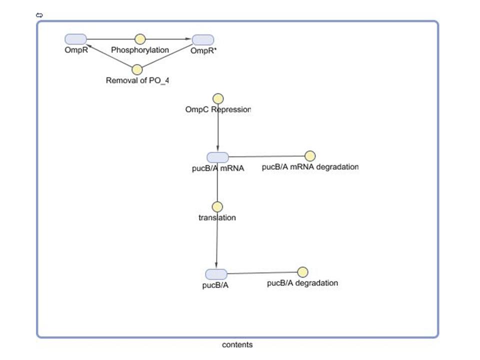
pucBA Expression Model Diagram
Modeling the Gene Regulatory Network
- Our group seeks to assess the optimality of the synthetic system that modulates pucB/A gene expression and LH2 complex assembly in Rhodobacter sphaeroides. Here we employ a mathematical model of this system to generate predictions about the behavior of the active system in response to light input. Features of the system that the model may help investigate include the time scale of response to light signals, the robustness of the system in response to fluctuations in light intensity, and the translation between changes in gene expression and the absorbance spectrum of the engineered cells.
- Though the context of the model can extend back to the transcription of PrrA/B genes involved in integration oxygen and light signals, a preliminary testing model was developed using assumptions of certain initial conditions to isolate the light signal's effect. Since Cph8 and OmpR are located on the same transcript downstream of the puc promoter region, it was assumed that their associated protein and mRNA had already reached steady state concentrations. Moreover, the concentrations of the factors were assumed to be equal at this state. The model whose diagram was constructed in the Simbiology Toolbox distributed by MathWorks details key reactions leading to the translation of the pucB/A genes. The reaction rate equation used for the phosphorylation of OmpR as a consequence of the light signal reaching Cph8 bound to OmpR is capture in a modified form of Michaelis-Menten kinetics. A logic function that corresponds to light ON/OFF (1/0) multiplies the maximum reaction rate in the numerator. Thus, the model assumes that no phosphorylation occurs by this mechanism in the absence of light. The OmpC promoter binding equation was based on the Hill Equation for a Repressor (1).
- Component characterization steps and literature searches are underway in order to obtain quantitative parameters for the reaction rates. In order to simulate behavior of the system, putative values were included that exaggerate true concentrations and time scales. OmpR was given an initial concentration normalized to one, and all other components were assumed insignificant initially to this value. An ideal light pulse was introduced at an instant and removed thirty simulation seconds later. From this rudimentary simulation it can be drawn that the nonlinearities of the phosphorylation and transcription factor binding kinetics effectively smooth the sharp light input. By design, the light switch ON yields phosphorylation of OmpR and repression of the pucB/A genes which would give rise to LH2. Conversely, when left OFF, the concentration of pucB/A recovers and increases until the steady state determined by its translation and degradation rates.
References
1. Alon, Uri. Introduction to systems biology and the design principles of biological networks. Boca Raton, FL: Chapman & Hall, 2006.
2. Bower, James M. Computational Modeling of Genetic and Biochemical Networks (Computational Molecular Biology). New York: M.I.T. PRESS, 2001.
3. System modeling in cellular biology from concepts to nuts and bolts. Cambridge, MA: MIT P, 2006.
Volver Arriba
 "
"


